FAQ’s for Koru Ultrasound & Care Centre
-
What are the opening times?
-
How do I book?
-
Do I require a referral from my Doctor?
-
What is Ultrasound imaging and how does it work?
-
What are the limitations of Ultrasound imaging?
-
What Ultrasound equipment do you use?
-
What should I wear for the examination?
-
What happens during the examination?
-
Is Ultrasound safe?
-
How long will my scan take?
-
Does the scan hurt?
-
When and how do I get the results?
-
Who interprets the results and how do I get them?
-
I am pregnant. What scans should I have and when?
-
How much does it cost?
-
How do I pay?
-
Are your staff qualified in the use of medical ultrasound equipment?
What are the opening times?
Please refer to opening hours on our home page.
How do I book?
Our friendly Receptionist will be able to book you an appointment time that suits you on 03 541 0050.
I am pregnant. What scans should I have and when?
First trimester: If you are very unsure of how many weeks pregnant you are you may be offered a dating scan to determine a more accurate due date. If you have had previous miscarriages or have a high risk of miscarriage you may also be offered an early scan. However, in many pregnancies, your first scan will be your nuchal translucency scan (NT scan) which happens between 11 weeks and 13 weeks and 6 days of pregnancy.
Second trimester: Another scan is done at about 20 weeks to check that your baby is developing normally (NSC 2007).
Third trimester: You may be recommended to have one or more growth scans between 28 weeks and 40 weeks if you've previously given birth to a small baby, if you are having twins or if you have other complications, for example, if you are diabetic. Your doctor or midwife may suggest a growth scan if your baby feels and measures smaller than expected.
Are your staff qualified in the use of medical ultrasound equipment?
Yes our Sonographers have post-graduate degrees in Clinical Ultrasound and have more than 25 years scanning experience between them. Our Radiologist, Brett Lyons, qualified in 1992 from Otago University.
You can read more about our staff here.
Do I require a referral from my Doctor?
For pregnancy scans a Midwife or GP referral is essential.
For other scans, including ACC scans, a referral is necessary from a GP or other appropriate Clinician, such as a Physiotherapist, Chiropractor or Osteopath.
When and how do I get the results?
Normally scan results will be sent to the referring Clinician within 24 hours. Sometimes provisional information (but not a clinical diagnosis) can be given at the time of the scan.
Who interprets the results and how do I get them?
A Radiologist (a Physician specifically trained to supervise and interpret radiology examinations) will analyse the images and send a signed report to the Clinician who requested the exam.
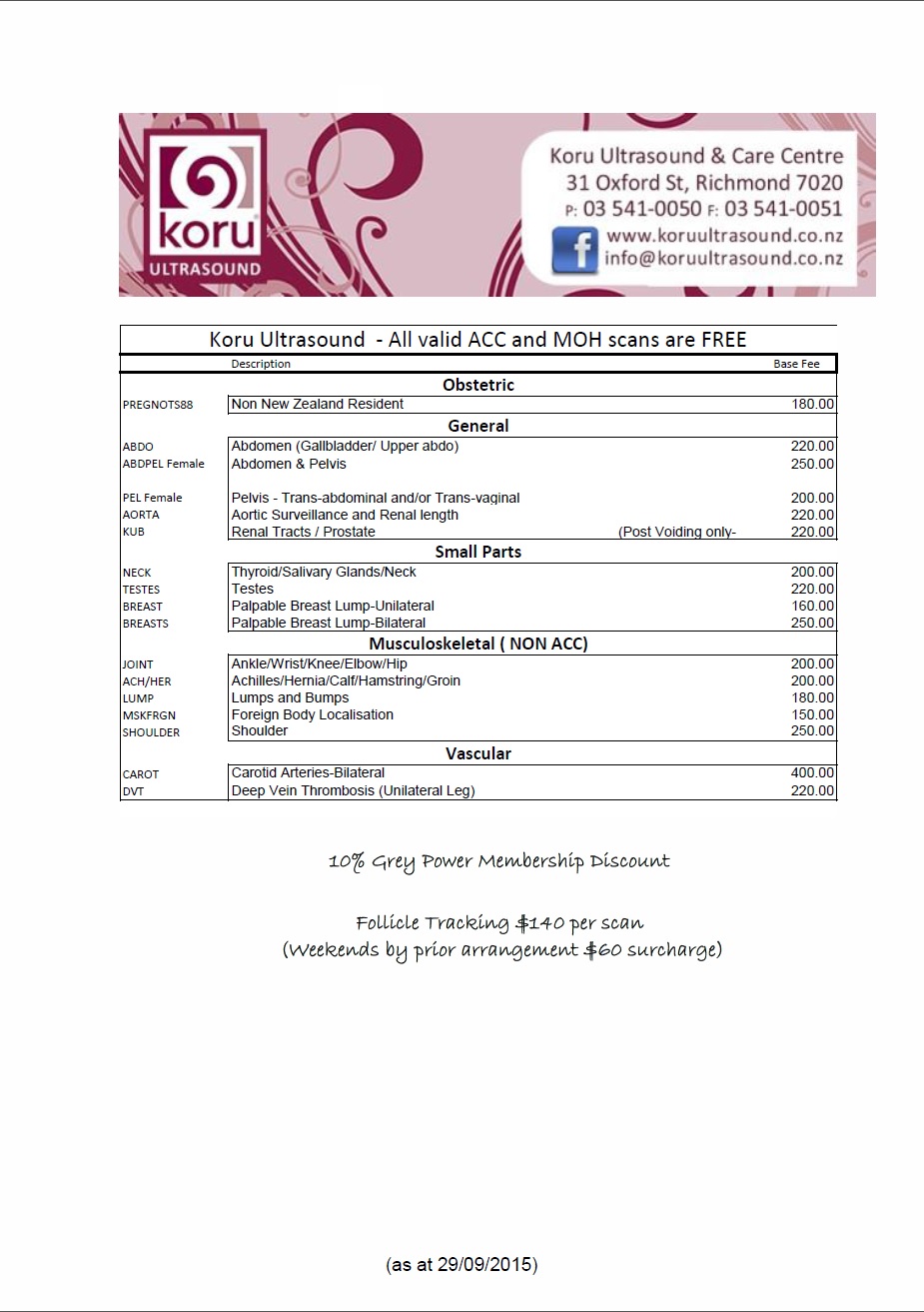
How much does it cost?
Current charges for all scans can be provided by our receptionist or by clicking here.
How long will my scan take?
The duration of the scan depends on its complexity, but is normally between 15 and 40 minutes.
How do I pay?
Where payment is necessary we prefer payment on the day of the scan by EFTPOS and/or credit card, but are also happy to accept payment by cash.
Does the scan hurt?
Ultrasound scans are completely painless.
Is Ultrasound safe?
Clinical ultrasound has been shown for many years to be safe.
What is ultrasound imaging and how does it work?
Ultrasound imaging is based on the same principles involved in the sonar used by bats, ships and fishermen. When a sound wave strikes an object, it bounces back, or echoes. By measuring these echo waves, it is possible to determine how far away the object is as well as the object's size, shape and consistency (whether the object is solid or filled with fluid).
In medicine, ultrasound is used to detect changes in appearance, size or shape of organs, tissues, and vessels or detect abnormal masses, such as tumours.
What are the limitations of ultrasound imaging?
Ultrasound waves are disrupted by air or gas; therefore ultrasound is not an ideal imaging technique for air-filled bowel or organs obscured by the bowel.
Large patients are more difficult to image by ultrasound because greater amounts of tissue weakens the sound waves as they pass deeper into the body.
Ultrasound has difficulty penetrating bone and, therefore, can only see the outer surface of bony structures and not what lies inside.
What Ultrasound equipment do you use?
Our ultrasound machine is a Philips IU22 High Resolution Scanner. In addition to its advanced imaging capabilities and easy to use features, the Philips iU22 helps to speed up the entire process, from imaging to data storage.
What should I wear for the examination?
You should wear comfortable, loose-fitting clothing for your ultrasound exam. You may need to remove clothing and jewellery in the area to be examined.
What happens during the examination?
For most ultrasound exams, you will be asked to lie face-up on an examination table that can be tilted or moved.
The Sonographer will apply a warm water-based gel to the area of the body being studied. The gel will help the transducer make secure contact with the body and eliminate air pockets between the transducer and the skin that can block the sound waves from passing into your body. The transducer is placed on the body and moved back and forth over the area of interest until the desired images are captured.
There is usually no discomfort from pressure while the transducer is pressed against the area being examined.
Once the imaging is complete, the clear ultrasound gel will be wiped off your skin. Any portions that are not wiped off will dry to a powder. The ultrasound gel does not stain or discolour clothing.
In a small proportion of ultrasound studies, the transducer is attached to a probe and inserted into a natural opening in the body. These exams include:
Transvaginal ultrasound: The transducer is inserted into a woman's vagina to view the uterus and ovaries. This application is used for early pregnancy confirmation (less than 8 weeks) and for improving the detail at times for Gynae scans.
On the occasions when this is recommended the process will be explained and permission from you will be sought.