 Women's Health ultrasound scans. Scans performed by women for women.
Finalist for the Nelson Tasman Chamber of Commerce Innovation award for our unique breast ultrasound density grading system and screening technique.
Phone us today for an appointment
(03) 541 0050
Breast Imaging
The current breast cancer screening method in NZ is 'breast awareness', until the age of 40 and then mammograms 2 yearly - which are only government funded between 45 and 69 years old (NSU).
For those outside of the funded age, or those known to have dense breasts from previous mammograms, please note that ultrasound provides additional options for screening (Leddy et al. 2013; Candelaria et al. 2013; .
You are given a breast density grading at your Koru Ultrasound appointment. Read more about Koru Ultrasound's breast density grading here.
More information here.
At Koru we offer 2 different kinds of breast ultrasound:
- Whole breast ultrasound screening for women with known or suspected dense breasts (both breasts)
- Targeted ultrasound of a breast lump (single breast)
Ultrasound is ideal for:
- All women with dense breasts
- Women under the age of 45 (frequently the density of breast tissue on younger women makes mammography less helpful)
- Pregnant or breast-feeding women
- Women with breast implants. We do not provide initial imaging Post surgery. Follow up implant ultrasound scans of both breasts are welcomed.
- Women who decline mammogram (30% of eligible women don't have regular mammograms)
- Women with a strong family history, who may wish to begin screening before they are eligible for free mammograms
- Women on Hormone Replacement Therapy (HRT) - who may also have increased breast density.
- Men with a palpable lump
All previous imaging reports are required on booking eg Mammogram/Thermogram/MRI.
Are Ultrasounds Scans safe?
Ultrasound uses sound waves, instead of radiation, so can be used as needed. Long worldwide experience of clinical ultrasounds has shown no adverse effects (FDA).
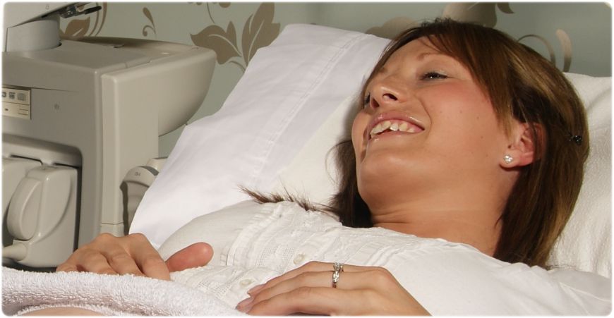
What happens when you have an Ultrasound?
The ultrasound specialist simply runs a probe over each breast in turn. The ultrasound specialist may use her hands to feel any lumpy areas that you or your doctor has identified. All of our ultrasound specialists and receptionists are female to help you feel at ease about the whole process. The images are made by thoroughly imaging the breast tissue in three planes: Radial (clock face), horizontal and vertical. Our radiologist then carefully examines the images and prepares a detailed report including breast density, which is available the next working day. It is common to find simple breast cysts and we prefer to scan you after your period if you are not menopausal, as breast tissue is less 'lumpy' at this time. We keep a 'breast map' of any cysts and their size, so that they can be monitored every year to exclude unusual growth.
Why Ultrasound?
Increased breast density reduces the chance of detecting a breast cancer on a mammogram (Girardi et al. 2013; Brem 2012). Ultrasound is better than mammography at looking at dense breast tissue and therefore is ideal for women with dense breasts, such as young women and many post-menopausal women, including those on Hormone Replacement Therapy (HRT) (Girardi et al 2013; Kornecki 2011; Brem 2012). Mammography can be a better screening tool for women who don't have dense breasts (Leddy et al. 2013;Girardi et al. 2013 ).
Ultrasound is a pain-free non-invasive method of examining the breasts. It can be used to evaluate several aspects of breast structure. The most common application is to determine the composition of a breast mass (Candelaria et al 2013; Kornecki 2011; O'Neill et al. 2012). The presence of large collections of calcifications may also be detected sonographically, but each collection or individual calcification must be fairly large, at least a few millimeters in diameter (Berg 2008; Candelaria et al. 2013; ).
Other applications include ruling out the presence of lymph node masses that often accompany breast cancer and evaluating the integrity of breast implants (Candelaria et al. 2013).
It is important to remember that ultrasound is often not capable of detecting the very small (approximately 1mm) calcifications that are visible with mammography and often represent the first signs of breast cancer.
Ultrasound is used as a complementary technique to mammography and is not a replacement.
FAQ
Q: I have been told at the age of 65 a breast screening by ultra sound is not effective due to breast density – is this correct?
A: No, a breast ultrasound is effective at any age, but is especially recommended in very young or breast feeding women as unlike mammography there is no ionising radiation.
Usually young women have dense breasts and as they get older they become more fatty, but this is not always the case. Some reports say 30- 40 % of women over 45 have dense breasts. Ultrasound is good at detecting problems regardless of breast density. Mammography is only good at picking up problems with fatty breasts and will not always pick up a tumour on a very dense breast. Mammography is quicker and cheaper to perform compared to a whole breast ultrasound and that is why it is funded by the government at an age when women are more likely to have fatty breasts, over 45. You cannot be sure of breast density by age or by looking/ palpating a breast, therefore a mammogram or ultrasound can be used to determine breast density. Woman are not always told their breast density after a mammogram, but we always give you a report of your breast density in our ultrasound reports
Q: How much does a breast ultrasound cost?
A: A single breast is $250 ( $160 with a community service card) Both breasts are $400 ($250 with a community services card)
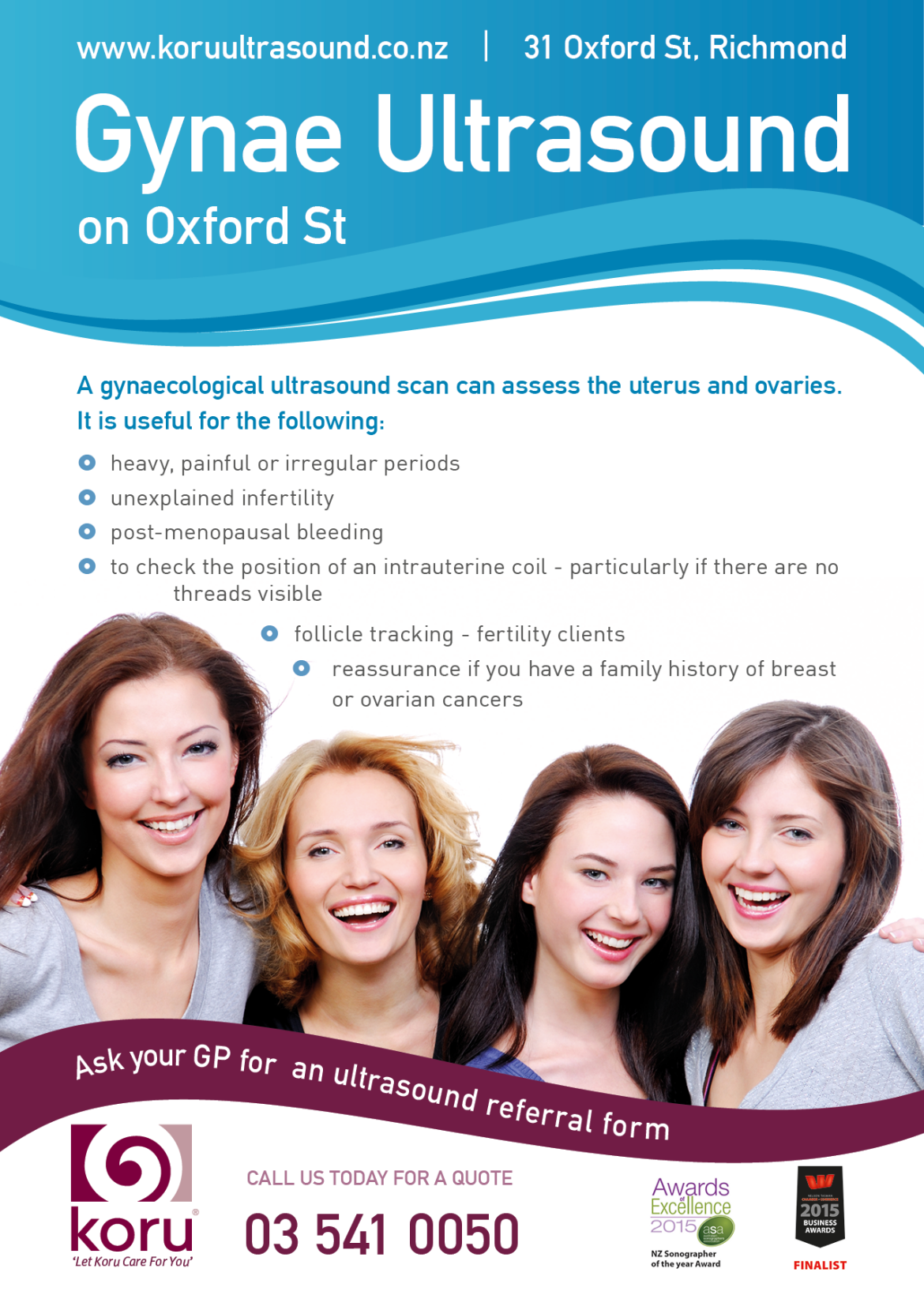
Gynaecological Imaging
Used to assess the uterus and ovaries. The types of problems which can be found on these scans include ovarian cysts, endometrial polyps and fibroids.
You need a full bladder for this scan. Please finish drinking 1L of water or other non-fizzy fluid 1hour before your appointment and do not go to the toilet.
It is likely that the sonographer (ultrasound specialist preforming the exam) will suggest preforming an internal vaginal scan to get closer to, and visualise, your internal organs better. This will help give a better diagnosis.
This should not be painful, and the scan is not harmful in any way. You will be asked to empty your bladder in our ensuite before this part of the examination.
IVF/Egg donation follicle tracking
Same day reporting of number and size of follicles for harvesting. Repeat scanning at 2 day intervals (including weekends) as needed.*